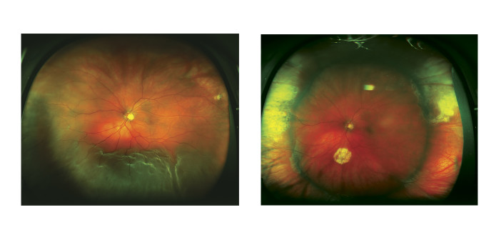
“Seeing floaters” is one of the most common complaints that I hear from patients. While the majority of us will experience at least one floater at some point in our lifetimes, it is very important to have a dilated eye exam if you begin to see new floaters in your field of vision, especially if they are associated with other symptoms, such as flashes of light.
What are floaters?
Floaters look like small dots or spider webs moving through your field of vision. Floaters can have different shapes and often look like strands, circles, clouds, or strings. They are easier to see on a bright day when you are looking at a light-colored background, like the sky.
What causes floaters?
The back of the eye is filled with a clear substance called the vitreous gel. As people reach middle age, the gel that was once solid beings to liquefy. As the gel liquefies, strands of proteins suspended in the gel can cast shadows on the retina, resulting in floaters. As the gel collapses, it can pull too hard on the retina, resulting in a tear that could lead to a retinal detachment if left untreated.
What are the symptoms of floaters?
While most floaters are not serious, you should pay attention to the following symptoms:
• One new, large floater or “showers” of floaters that appear suddenly
• Seeing sudden, repeated flashes of light like lightening or camera flashes, even when your eyes are closed
• Vision loss in one eye
• A shade or curtain coming down over your field of vision
If you experience any of these symptoms, you should call your eye doctor to schedule a dilated eye exam.
How are floaters treated?
Generally, treatment is not recommended for floaters. Though they can be bothersome in certain lighting conditions, they generally do not result in loss of vision. However, new floaters, especially if associated with flashes of light, could mean that the vitreous gel has pulled too hard on the retina as it liquefies. If your doctor finds a tear or break in the retina, they may refer you urgently to a retina specialist for repair of the retinal tear, or even surgery if you have developed a retinal detachment.
What is a retinal detachment?
If you compare the eye to a camera, the retina is the inner layer of the back of the eye and functions like the film in a camera. Light entering the eye is focused on the retina, resulting in an image that is sent to the brain to provide vision. If the retina is torn, fluid from within the eye can travel through the tear and lift the retina away from the wall of the eye, resulting in a retinal detachment. If this happens, you may experience a progressive shadow or curtain in your vision that may even involve your central vision. A detached retina is a very serious condition that can lead to permanent vision loss.
How are retinal detachments treated?
Generally, a retinal detachment requires surgery to repair. The timing of surgery depends on the nature of the detachment, and a delay in diagnosis can increase the risk of vision loss. There is a variety of methods that your surgeon can use to repair the retina, including some combination of a vitrectomy, scleral buckle, and/or a gas bubble (pneumatic retinopexy). Often, these treatments are done in combination with laser or freezing treatment (cryopexy) to seal the retinal tears. In addition, gas or silicone oil are commonly used to hold the retina in place while it heals.
Summary
Overall, floaters are a common symptom of natural, age-related changes in the gel of the eye. However, in some cases, floaters can be a sign of a more serious eye condition, such as retinal tears or retinal detachment. While these conditions can usually be successfully repaired, timely diagnosis can protect you from vision loss. Be sure to call your eye doctor if you experience any of the symptoms above.


