
Sometimes, the smallest efforts can make the biggest difference. In terms of random acts of kindness this is certainly true, and recently, with applied research at Kettering University, small – or, more accurately –“nano-sized” efforts will eventually yield substantial results.
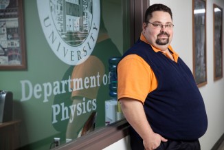
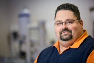
Physics professor, Dr. Ron Tackett is contributing to the continuous battle against cancer; but he and a few other professors at Kettering are attempting to conquer it from the smallest angle by using magnetic nanoparticles as a non-invasive treatment for certain types of cancer.
“Very rarely, an undergraduate institution can get log-on time with an instrument like the TEM. We see it as our niche to fill.”
Dr. Ron Tackett

Since 2013, the Kettering faculty has secured more than $2 million in equipment through Major Research Instrumentation (MRI) grants from the National Science Foundation (NSF). This has been Kettering’s eighth award from the NSF since 2012 – the most of any institution of higher education in Michigan.
With this grant, Dr. Tackett and other members of the Kettering staff are anticipating the delivery of a high-resolution Transmission Electron Microscope (TEM) in the next couple of months. This microscope will allow them to take high-resolution pictures of magnetic nanoparticles. In addition to some assistance from other students, Dr. Tackett has been collaborating with other professors in this research: Dr. Prem Vaishnava, Dr. Ronald Kumon, Dr. Corneliu Rablu, and Dr. Lihua Wang.


 Working with various neurosurgeons in Flint, Dr. Tackett has gathered information on certain tumors that will be positively affected by this research – tumors like glioblastoma multiformae. Glioblastoma tumors are the most common brain tumors, growing in tendrils, so even if doctors go in surgically, it is difficult to remove it all. Currently, there is not a cure for glioblastoma; those tumors don’t respond well to chemotherapy or other treatments. This nanotechnology will make the tumors easier to remove by essentially “burning up” and killing the tumor, a treatment less invasive to the human body.
The researchers’ goal is to make the particles as magnetic as possible, but still biocompatible. If they are successful, treatment for patients would not take very long. It’s possible that it could be a one-day procedure. “There’s no reason to believe that the patient wouldn’t just walk away from this,” Dr. Tackett believes.
Even if the tumor is not completely destroyed by the heat, after the treatment, the tumor is much more sensitive to radiation for a certain period of time. Radiation more effective, using lower doses, less invasive. “The goal is to treat this cancer in a much more humane way,” Dr. Tackett says.
And this goal will most likely be accomplished through heating up the tumors with these magnetic nanoparticle injections. The nanoparticles, from a physics standpoint, will absorb energy from the field, or appear to be dancing and vibrating. This technique will locally target these cancerous tumors without doing damage to surrounding tissue. “The body can flush these nanoparticles, hopefully, because they’re so small,” Dr. Tackett adds. These nanoparticles are tens to hundreds of nanometers in diameter; one nanometer is a billionth of a meter – smaller than a human hair – and is not visible to the naked eye.
The reason physicists like Dr. Tackett are pursuing this type of cancer in their research is purely because there are not many treatments for it. It is believed that a similar method can be used with retinoblastoma (eye cancer), because that cancer is also not easily treated. Extremity-based cancers, like breast cancer, are the best candidates to benefit from this treatment. Any cancers in the torso are not good candidates because the heart (and other vital organs) should not be heated by magnetic nanoparticles.
 Luckily, with Kettering acquiring the Transmission Electron Microscope through the NSF-MRI grant, immediate results are expected.
A TEM is nothing like a run-of-the-mill microscope, according to Dr. Tackett. Scheduled to arrive in 2016, the TEM is the only scientific instrument that can see nanoparticles. It uses electrons to see images, rather than light. The shorter wavelengths of the electrons result in better resolution (down to a quarter of a nanometer). In some pictures, they can see down to the atom level. Determining the shapes, sizes and distribution of the nanoparticles will be the most helpful to the research; once they discover the nature of the particles, it will help them understand how to make them as magnetic as possible, thus having better results with removing glioblastoma tumors. The nearest TEM is at Wayne State University, where Dr. Tackett occasionally does some of his research.
For teaching students at Kettering, it will open many new doors in the minds of students of physics and other areas to wrap their heads around complex research. “A TEM is a staple instrument in the field of material science,” says Dr. Tackett. “It’s my vision to have a 60- or 70-inch TV screen showing what’s on the monitor for the TEM, so a group of students can see it. They could start visualizing it right there.” The TEM, along with the cancer research, is continuing to make Kettering University stand out as the prime institution for those in the applied sciences. “Very rarely, an undergraduate institution can get log-on time with an instrument like this,” Dr. Tackett says. “We see it as our niche to fill.”
Photography by Mike Naddeo


